
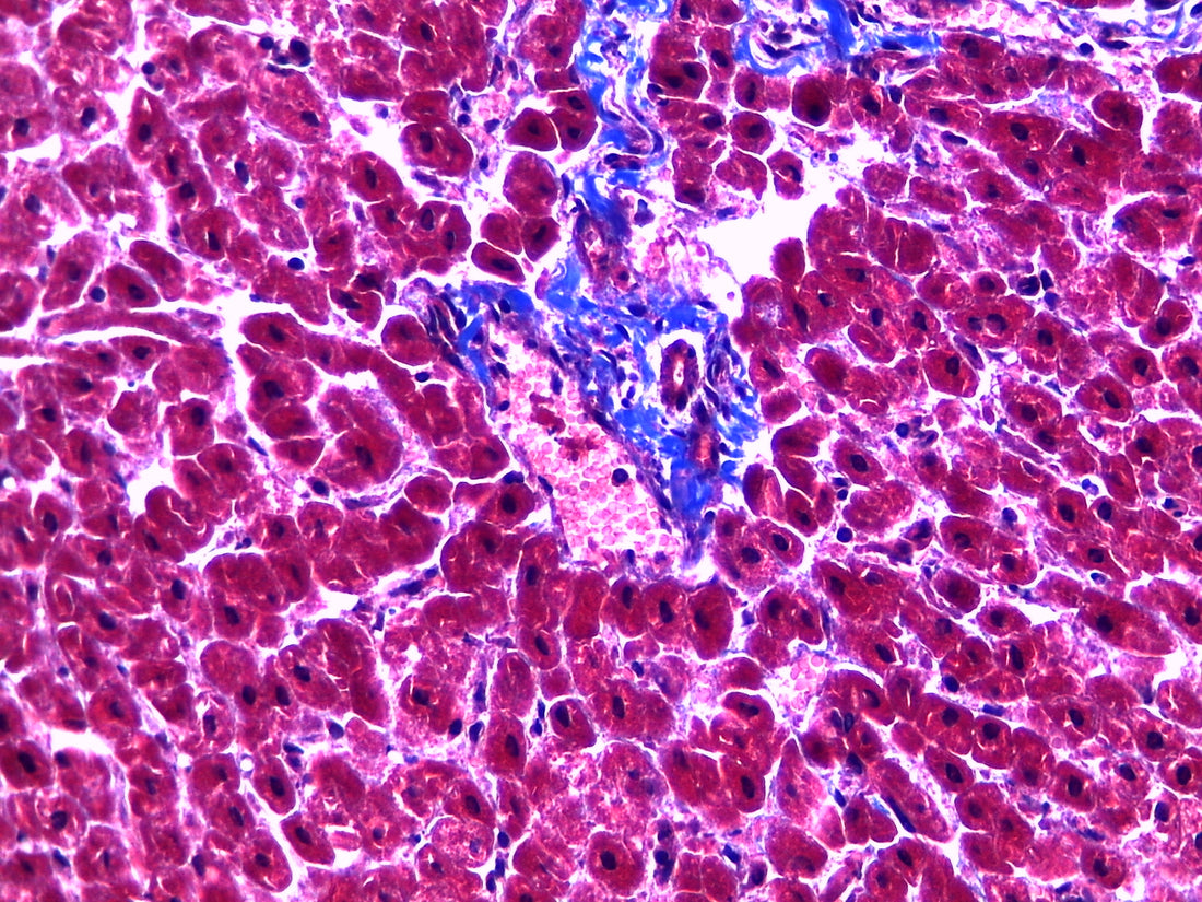
Masson's trichrome stain is a histological staining technique used to differentiate and visualize various tissue components, particularly collagen fibers, muscle fibers, and erythrocytes. The stain is named after the French pathologist Raymond Masson who developed the technique in the early 1900s.
Under the microscope, nuclei appear dark blue, cytoplasmic structures such as muscle fibers and erythrocytes appear red, and collagen fibers appear blue-green. This allows for the visualization and differentiation of different tissue components.
Masson's trichrome stain is commonly used in the diagnosis of various diseases such as myocardial infarction, liver cirrhosis, and fibrosis. It is also used in research to study tissue remodeling and fibrosis.
